Thursday, December 7, 2006

Patient: Khong Fay Fay

Patient Name: Khong Fay Fay
Sex: Female
Age: 26 years old


Urinary tract infections
The urinary tract is divided into:
Lower portion - urinary bladder and the urethra.
Upper portion - kidneys, renal pelves, and ureters
Upper urinary tract infections (UTIs) are most commonly ascending; they originate in the urinary bladder and ascend through the ureters to the kidneys.

The symptoms of upper UTIs are fever (often with chills) and flank pain. Frequency, urgency, and dysuria are more suggestive of infections of the urinary bladder and urethra (Lower urinary tract).

Community acquired UTI: Cystitis
Cystitis is caused bladder mucosal invasion, most often by enteric coliform bacteria (eg, Escherichia coli) that inhabit the periurethral vaginal introitus and ascend into the bladder via the urethra. Sexual intercourse may promote this migration, and cystitis is common in otherwise healthy young women.

http://www.emedicine.com/EMERG/topic626.htm

Community acquired UTI: Acute Pyelonephritis
Pyelonephritis is an ascending urinary tract infection that has reached the pyelum (pelvis) of the kidney. The causes are lower UTIs, mainly cystitis and prostatitis (but in this case, a woman does not have a prostate gland)

Williams DH, Schaeffer AJ (2004). "Current concepts in urinary tract infections". Minerva Urol Nefrol 56 (1): 15-31.


Discussion
The symptoms of upper and lower UTIs are relatively similar. Absence of catherter insertion inidcates that the UTI was not acquired in hospital. Thus the area of focus will be on community acquired UTIs. With consideration of the patient’s age and sex (26 years old female), it is highly probable that she is suffering from cystitis or acute pyelonephritis. In addition to the female urethra being shorter and nearer to the anus (anatomy), the fact that young women who are sexually active (26years old) will be more prone to cystitis (or communtiy acquired UTIs)

Small list of the common microbes related to UTI:
Escherichia coliCommon in young women, causes 80% of community-acquired UIT and 90% of the urinary tract infections (UTI) in anatomically-normal, unobstructed urinary tracts.

Staphylococcus saprophyticus - In young women, S. saprophyticus is, after Escherichia coli, the second-most-frequent causative agent of acute UTI

Proteus mirabilis - with functional or structural abnormalities or with long-term catheterization, forms bladder and kidney stones as a consequence of urease-mediated urea hydrolysis.

Pseudomonas aeruginosa - usually hospital-acquired and related to urinary tract catheterization, instrumentation or surgery


http://textbookofbacteriology.net/e.coli.html
http://iai.asm.org/cgi/content/abstract/72/5/2922
http://textbookofbacteriology.net/pseudomonas.html
Hovelius B, Mardh PA. (1984) “Staphylococcus saprophyticus as a common cause of urinary tract infections”. Rev Infect Dis. May-Jun; 6 (3): 328-37

Highly suspected microbes: Escherichia coli and Staphylococcus saprophyticus.


Tests
Dipstick tests
Dipstick tests are rapid and inexpensive, but need to be interpreted with caution. The two commonly used tests are the:

Leukocyte esterase test
Indicates pyuria by detecting esterases released from white blood cells. However, pyuria is not a specific indicator of infection.

Nitrate reduction test
Detects nitrites produced from nitrates by bacteria (mainly Gram-negative bacteria).
Dipstick tests are of most use as a negative screen


Urine cultures (subjected to overnight incubation at 37 degrees Celsius)

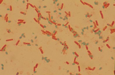
MacConkey Agar - mainly used in identification of lactose fermenting, Gram-negative enteric pathogens and for inhibiting growth of Gram-positive organisms. Bacterial colonies that can ferment lactose turn the medium red. This red color is due to the pH indicators response to the acidic environment created by fermenting lactose. Organisms that do not ferment lactose do not cause a color change.


Catalase test - The catalase test involves adding hydrogen peroxide to a culture sample or agar slant. If the bacteria in question produce catalase, they will convert the hydrogen peroxide and oxygen gas will be evolved. The evolution of gas causes bubbles to form and is indicative of a positive test.


Coagulase test
- for differienting between pathogenic and non-pathogenic strains of Staphylococcus. A positive test is denoted by a clot formation in the test tube after the allotted time.


Indole test - When tryptophan is broken down, the presence of indole can be detected through the use of Kovacs' reagent. Kovac's reagent, which is yellow, reacts with indole and produces a red color on the surface of the test tube.


Oxidase test
- If the bacteria oxidize the disk (remove electrons) the disk will turn purple, indicating a positive test. No color change indicates a negative test

http://medic.med.uth.tmc.edu >path >tests


Flow chart to direct the identification of serveral bacterial. (Main focus is on E. Coli)


A flow chart for the identification of Gram-Positive bacteria. (Staphylococcus saprophyticus not shown) Staphylococcus saprophyticus has the same characteristics as S. epdermidis, but Staphylococcus saprophyticus is resistant to novobiocin.

Use of Novobiocin-containing medium (sensitivity disks with 5 micro grams of novobiocin)

By demonstrating the resistance to the anti-microbial agent novobiocin, staphylococcus saprophyticus can be identified amongst the coagulase-negative staphylococcus.

http://web.indstate.edu/thcme/micro/staph/sld003.htm

Robert H. Latham, Grada A. Grootes-Reuvecamp, Dolores Zeleznik, and Walter E. Stamm. (1983). "Use of a Novobiocin-Containing Medium for Isolation of Staphylococcus saprophyticus from Urine." J Clin Microbiol. June; 17(6): 1161–1162.

-Willie-

Urinary tract infection


Patient 3
Name: Maisy Wong
Sex: Female
Age: 66 yrs
Complaints: Fever, chills, bladder distension, on indwelling catheter
Diagnosis: Urinary tract infection
Antibiotic treatment: Nil

Maisy Wong is on indwelling catheter. An indwelling urinary catheter is a tube that drains urine from the bladder into a bag. The tube is placed into the urethra (the part of the body that drains the bladder) and up into the bladder. Catheter-associated urinary tract infections are caused by a variety of pathogens, including Escherichia coli, Klebsiella, Proteus, enterococcus, Pseudomonas, Enterobacter, Serratia, and Candida. Many of these microorganisms are part of the patient's endogenous bowel flora, but they can also be acquired by cross-contamination from other patients or hospital personnel or by exposure to contaminated solutions or non-sterile equipment. Catheter-associated urinary tract infections are generally assumed to be benign. Such infection in otherwise healthy patients is often asymptomatic and is likely to resolve spontaneously with the removal of the catheter.

Microbiological Media Used
Blood Plate Agar (BAP)

Contains blood from a mammal (usually sheep), and respires to typical transperent nature, typically at a concentration of 5–10%. BAP are an enriched, differential media used to isolate fastidious organisms and detect hemolytic activity. β-hemolytic activity will show complete lysis of red blood cells surrounding colony, while α-hemolysis will only partially lyse hemoglobin and will appear green. γ-hemolysis is the term referring to a lack of hemolytic activity.


Cysteine Lactose Electrolyte Deficient Agar (CLED)
Is a valuable non-inhibitory growth medium used in the isolation and differentiation of urinary organisms. Being electrolyte deficient, it prevents the swarming of Proteus species. Lactose fermenters produce yellow colonies on CLED agar; non-lactose fermenters appear blue.

Most probable microorganisms:
Proteus mirabilis
Pseudomonas aeruginosa
E.coli
http://www.cdc.gov
http://www3.umdnj.edu

Posted by Xiu hui


Microorganisms Suspected
Patient 2: Kwan Siew Lan

1) Salmonella typhi

Description
- Facultative anaerobes
- Gram-negative rods
- Non-lactose fermentors
- Produce H2S
- Causes infection in the lining of the small intestine


Picture of Salmonella typhi taken from (http://www.nlm.nih.gov/ >
Medline > Medical Encycopedia >S-Sh > Samonella Enterocolitis)

Causes and Risk Factors
- Ingestion of contaminated food or water
- Ingestion of improperly prepared or stored food (especially undercooked turkey or chicken, unrefrigerated turkey dressing, undercooked eggs)
- Family members with recent salmonella infection
- Recent family illness with gastroenteritis
- Institutionalization
- Recent poultry ingestion
- Owning a pet iguana or other lizards, turtles, or snakes (reptiles are carriers of salmonella)
- Old or young age
- Patients with impaired immune systems
(http://www.nlm.nih.gov/ > Medline > Medical Encycopedia > S-Sh > Samonella Enterocolitis)

Transmission
- Transmitted mainly via fecal-oral rout and food prepared by chronic carriers

Signs and Symptoms
- Abdominal pain or cramping or tenderness
- Mild to severe diarrhea
- Nausea
- Vomiting
- Fever
- Chills
- Muscle pain
(http://www.nlm.nih.gov/ > Medline > Medical Encycopedia > S-Sh > Samonella Enterocolitis)


2) Campylobacter jejuni

Description
- Gram-negative “S” or “gull wing” shaped
- Motile with a single polar flagellum
- Micro-aerophillic (5%O2 with 10%O2)


Picture of Campylobacter jejuni taken from (http://www.nlm.nih.gov/ >
Medline > Medical Encycopedia > C-Cg > Campylobacter Enteritis)

Causes and Risk Factors
- Eating or drinking contaminated food or water, often raw poultry, fresh produce, or unpasteurized milk
- Close contact with infected people or animals
- Recent travel in an area of poor hygiene or sanitation
(http://www.nlm.nih.gov/ > Medline > Medical Encycopedia > C-Cg > Campylobacter Enteritis)

Transmission
- Via Faecal-oral route, contaminated food and water with animal faeces

Signs and Symptoms
- Cramping abdominal pain
- Watery diarrhea, sometimes bloody
- Fever
(http://www.nlm.nih.gov/ > Medline > Medical Encycopedia > C-Cg > Campylobacter Enteritis)

3) Vibrio parahaemolyticus

Description
- Curved, comma-shaped gram-negative rods
- Highly motile with polar flagella
- Grow well at alkaline pH (8.5-9.5)
- Halophillic bacteria that grow well in high NaCl concentration

Picture of Vibrio parahaemolyticus taken from

Causes and Risk Factors
- Eating raw or undercooked shellfish, particularly oysters
- Infection in the skin when an open wound is exposed to warm seawater
- Persons with weakened immune systems
(http://www.cdc.gov > Diseases and Conditions > V > Vibrio parahaemolyticus Infection)

Signs and Symptoms
- watery diarrhea with abdominal cramping
- nausea
- vomiting
- fever
- chills
(http://www.cdc.gov > Diseases and Conditions > V > Vibrio parahaemolyticus Infection)


REASONS
Samonella typhi, Campylobacter jejuni and Vibrio parahaemolyticus commonly cause enterocolitis or some form of infection in the intestine with the symptom of diarrhea of varying degrees.
Posted by Melva
The microbe that I highly suspected are listed below:
Bacteria


Virus


www.aggie-horticulture.html
Plates used
· Blood plate agar (BAP): Blood Plate agar contains blood from a mammal (usually sheep), and respires to typical transperent nature, typically at a concentration of 5–10%. BAP are an enriched, differential media used to isolate fastidious organisms and detect hemolytic activity. β-hemolytic activity will show complete lysis of red blood cells surrounding colony, while α-hemolysis will only partially lyse hemoglobin and will appear green. γ-hemolysis is the term referring to a lack of hemolytic activity.

· Salmonella-Shigella Agar modified (SS): Beef Extract, Enzymatic Digest of Casein, and Enzymatic Digest of Animal Tissue found in SS plate provide sources of nitrogen, carbon, and vitamins required for organism growth. Lactose is the carbohydrate present in Salmonella Shigella Agar. Bile Salts, Sodium Citrate and Brilliant Green inhibit Gram-positive bacteria, most coliform bacteria, and inhibit swarming Proteus spp., while allowing Salmonella spp. to grow. Sodium Thiosulfate and Ferric Citrate permit detection of hydrogen sulfide by the production of colonies with black centers. Neutral Red is the pH indicator.

· Campylobacter Selective medium: A blood free medium, which will support the growth of enteric Campylobacter species. The selective supplements cefaperazone and amphotericin make the medium selective for Campylobacter jejuni and Campylobacter laridis when incubated at 37°C. Incubation at 42°C is no longer necessary and higher recovery rates have been reported at 37°C. Blood is replaced in the medium with charcoal, ferrous sulphate and sodium pyruvate, which enhance the growth and aerotolerance of Campylobacter species.

· MacConkey Agar (MAC): MacConkey agar is a differential plating medium recommended for use in the isolation and differentiation of lactose-fermenting organisms from nonfermenting Gram-negative enteric bacteria. It is selective by the presence of specific inhibitors.

· Thiosulphate-citrate bile sucrose agar (TCBS): A selective isolation medium for pathogenic Vibrio species. Most Enterobacteriaceae other than Vibrio species are suppressed for at least 24h. Bile salts inhibit Gram-positive organisms. Sodium thiosulphate serves as a source of sulphur, which, in combination with ferric citrate, detects hydrogen sulphide production. When sucrose is fermented it produces acid changing the pH. This is indicated by bromothymol blue and thymol blue. The medium is also alkaline which enhances the recovery of Vibrio cholerae.

· Selenite broth: Selenite Broth contains enzymatic digest of casein and enzymatic digest of animal tissue that provides nitrogen and vitamin sources. The main carbohydrate that is present in the broth is lactose. Lactose is the fermentable carbohydrate. Sodium Phosphate in the broth acts as a buffer. A rise in pH decreases selective activity of Selenite. The acid produced by lactose fermentation helps to maintain a neutral pH. Sodium Selenite inhibits the growth of Gram-positive bacteria and many Gram-negative bacteria.

· Alkaline peptone water (APW): Alkaline Peptone Water is an enrichment medium used for the cultivation of Vibrio species from feces and other infected materials. Clinical materials containing small numbers of Vibrio should be inoculated into an enrichment medium prior to plating onto a selective medium, such as TCBS Agar. Alkaline Peptone Water is a suitable enrichment broth for this purpose. The relatively high pH of the medium (approximately 8.4) provides a favorable environment for the growth of vibrios.

· Cefsulodin-Irgasan-Novobiocin (CIN) agar for Yersinia enterocolitica: Cefsulodin-Irgasan-Novobiocin (CIN) agar is a differential and selective medium for the isolation of Yersinia enterocolitica. Fermentation of mannitol in the presence of neutral red produces characteristic "bull's-eye" colonies. These are colourless with a red centre. A zone of precipitated bile may also be present. Crystal violet, sodium desoxycholate, cefsulodin, Irgasan (triclosan) and novobiocin are inhibitory agents.

· Sorbitol MAC for enterohaemorrhagic E.coli: Sorbitol MacConkey Agar medium contains sorbitol instead of lactose and it is recommended for the detection of enteropathogenic strains of E. coli, which ferments lactose, but does not ferment sorbitol and hence produce colorless colonies. Sorbitol fermenting strains of E. coli 0157:H7 produce pink-red colonies. The red colour is due to production of acid from sorbitol, absorption of neutral red and a subsequent colour change of the dye when pH of the medium falls below 6.8.

· Loeffler’s methylene blue: This solution is a solution that can be used alone as a simple stain, positive stain or as the counterstain in the acid fast stain procedure (Ziehl-Nielsen). It can also be used in the staining procedure that detects metachromatic granules (volutin).

· Enterococcosel Agar with 6 microgram / ml of vancomycin (VRE): Enterococcosel Agar Enterococcosel Agar incorporates Bile Esculin Azide Agar to yield rapid, selective detection and enumeration of enterococci. The surveillance for Vancomycin-Resistant Enterococci (VRE) can be accomplished by plating stool cultures onto Enterococcosel Agar with Vancomycin (6 µg/mL).


Microscopy
A wet preparation is examined for the presence of leucocytes and erythrocytes. Their presence may indicate invasive disease. This test is only done on request as its usefulness is limited.

1. Place a drop of liquid faeces or saline suspension of the faecal specimen on a microscope slide. Any mucous or flecks of pus or blood that may be present should be included in the suspension as these are likely to harbour disease causing organisms
2. Mix 1 drop of Loeffler’s methylene blue stain with the faeces specimen. Note that there must be an equal volume of faeces to stain.
3. Place a cover slip over it.
4. Wait 2 – 3 minutes for the nuclei to stain and then read the preparation under high power on (40x)
5. Observe for predominating numbers of white blood cells (WBCs), which indicate an invasive pathogen.

Culture
Inoculated places are O2 incubated unless otherwise indicated in the table.

All faeces are inoculated onto the following media: BAP, MAC, SS, Selenite broth and Campylobacter selective agar for the isolation of salmonella, Shigella and Campylobacter spp.

In addition to the above:
· Bloody faeces are also inoculated onto Sorbitol MAC plate.
· Watery faeces are plated on TCBS and inoculated onto APW to culture for Vibrio spp.


After culturing, gram staining is performed to differentiate gram positive (purple) and negative (pink). Gram negative bacteria includes gram negative cocci, bacilli and coccibacilli while positive includes gram positive cocci and bacilli.


posted by huiling

Wednesday, December 6, 2006

Patient 5

Patient 5
Name: Wong Wei Hong
Sex: Male
Age: 67 years
Complaints: Fever, chills, bladder distension; on indwelling catheter
Diagnosis: Urinary Tract Infection
Antibiotic treatment: Nil
Specimen: Urine

Definition
Urinary Tract Infection, or known as UTI, is an infection affecting one or more components of the urinary tract, which consists of two kidneys, two ureters, a bladder and a urethra.

Cause of Infection
A possible way for this patient to be infected with UTI is through the insertion of the indwelling catheter into his bladder. The purpose of inserting the indwelling catheter is to allow the drainage of urine out of the bladder. This would indicate to us that the patient might be suffering from a kidney disorder, as he cannot empty his bladder.

However, the use of a catheter may physically disturb the protective lining of the bladder wall, thus allowing bacteria to invade the exposed epithelium In addition to this, when the catheter is not thoroughly sterile, this could cause microorganisms to be present on the surface of the catheter upon insertion into the bladder. This would allow the bacteria to proliferate inside the bladder and thus resulting in the patient to develop a UTI infection.

Investigation Required
Urinalysis
It involves a physical and chemical examination of urine. In addition, the urine is spun in a centrifuge to allow sediments containing blood cells, bacteria, and other particles to collect. This sediment is then examined under a microscope. A urinalysis offers a number of valuable clues for an accurate diagnosis by,

• Observing the colour of the urine and presence of cloudiness (if any)
• Measuring the acidity of the urine
• Counting the number of leukocytes, which is indicative of UTI


Urine Culture
The procedure to detect for any bacterial growth in the urine is called a urine culture. This method involves the inoculation of the urine specimen onto agar mediums. The mediums required are Blood Agar, MacConkey Agar and Cysteine Lactose Electrolyte Deficient (CLED) Agar. The inoculated specimen is then streaked on the respective plates.

Blood Agar contains general nutrients and 5% sheep blood and it is useful for cultivating fastidious organisms and for determining the hemolytic capabilities of an organism.

MacConkey Agar is a differential medium used to isolate and distinguish lactose-fermenting organisms from non-fermenting Gram-negative enteric bacteria.

CLED Agar is a selective medium and inhibits swarming of Proteus species. In addition to this, they promote the growth of Candida and prevent the growth of pseudomonal.

All the 3 agar mediums will then be placed in an O2 incubator and left overnight before they are read the following day. If the Blood Agar shows a ‘swarming’ effect, this would probably indicate that the microorganism is Proteus mirabilis.

Gram Staining
After a urine culture has been performed, a Gram stain would be done using the isolated colonies from any of the agar mediums. This would help to identify if the microorganism is a Gram-positive cocci (GPC) or a Gram-Negative bacilli (GNB). Under microscopy, a GPC would appear bluish or purplish in colour and the cells would be circular. On the other hand, a GNB would turn out reddish or pinkish in colour while the cells would appear rod-shaped.

Biochemical Tests
After Gram staining has been, a few biochemical tests would be done to determine the suspected microorganism. A GNB would normally be an Escherichia Coli (E. coli) and to prove this, an oxidase test is done. E. coli would turn out to be oxidase negative. A GPC would most likely turn out to be a staphylococcus species, and a microorganism under this species would be Staphylococcus aureus. And to determine if the microorganism is likely to be Staphylococcus aureus, a latex test is done. This micro organism would be latex positive.

Antibiotic Susceptibility Test
After the microorganism has been classified, an antibiotic susceptibility test would be carried out to determine the antibiotics that the microorganism is sensitive to. This would allow the physician to prescribe the necessary antibiotics to the patient. In the event that the microorganism is resistant to majority of the antibiotics, stronger types of antibiotics would be given.

Suspected Organisms
Some of the suspected organisms that could lead to a UTI infection are listed as follows.

1) Escherichia coli
2) Proteus mirabilis
3) Pseudomonas aeruginosa
4) Staphylococcus aureus


References

http://www.reutershealth.com/
http://calder.med.miami.edu/
http://dentistry.ouhsc.edu/
http://en.wikipedia.org


Posted by Farhan

Thursday, November 30, 2006

Patient 4: Ng Ming En
Diagnosis: Food Poisoning
Complaints: Severe vomiting, diarrhea, abdominal cramps

Findings:
Foodborne illness (food poisoning) results from eating food contaminated with bacteria (or their toxins) or other pathogens such as parasites or viruses.
After they are swallowed, there is a delay, called the incubation period, before the symptoms of illness begin. This delay may range from hours to days, depending on the organism, and on how many of them were swallowed. During the incubation period, the microbes pass through the stomach into the intestine, attach to the cells lining the intestinal walls, and begin to multiply there. Some types of microbes stay in the intestine, some produce a toxin that is absorbed into the bloodstream, and some can directly invade the deeper body tissues. The symptoms produced depend greatly on the type of microbe. Numerous organisms cause similar symptoms, especially diarrhea, abdominal cramps, and nausea.

Culture


To find out what is the organism, the specimen (stools) is send for culturing using agar plates.
Inoculated places are O2 incubated unless otherwise indicated.

All faeces are inoculated onto the following media: BAP, MAC, SS, Selenite broth and Campylobacter selective agar for the isolation of salmonella, Shigella and Campylobacter spp.

In addition to the above:
· Bloody faeces are also inoculated onto Sorbitol MAC plate.
· Watery faeces are plated on TCBS and inoculated onto APW to culture for Vibrio spp.

Common Bacteria found in Bacterial gastroenteritis


Then gram staining is performed to determine whether organism is gram positive or negative. Following that, the organism shape (cocci or bacilli) is also determined and biochemical test is determined.the biochemical testsare used to detect the presence of enzymes (eg. Catalase, oxidase etc) and metabolic end-products (eg. Methyl red, etc). Lastly, an antibiotic sensitivity test is performed.

The above steps are performed to determine the organism and the organism I suspected is:Bacillus cereus
Some info about Bacillus Cereus
Bacillus Cereus food poisoning is a gastrointestinal intoxication caused by toxins produced by the Bacillus Cereus bacteria. It is a gram-positive spore-forming organism found in soil and dust. It is frequently found in rice dishes, occasionally pasta, meat or vegetable dishes, dairy products, soups, sauces and sweet pastry products where these have not been cooled quickly and effectively after cooking and during storage. It causes two different forms of food poisoning: an emetic illness and a diarrhoeal illness. The emetic illness ismediated by a highly stable toxin that survives high temperatures and exposure to trypsin, pepsin and pH extremes. The diarrhoeal illness is mediated by a heat- and acid-labile enterotoxin.

B. cereus-associated foodborne illness occurs as 2 distinct syndromes: emetic and diarrhoeal.
Incubation: Emetic; 1-6 hours after eating contaminated food.
Diarrhoeal; 10-12 hours.
Symptoms: The symptoms of the emetic syndrome result from ingestion of pre-formed toxin: nausea and vomiting, occasionally followed by diarrhoea. Diarrhoeal symptoms results from ingestion of vegetative organisms or spores and their subsequent multiplication and toxin production within the intestinal tract: abdominal pain, watery diarrhoea and occasional nausea

a picture of bacillus cereus
Another organism that i suspect in this patient is: Vibrio cholerae
posted by Huiling
ENTEROCOLITIS

Patient 2: Kwan Siew Lan
Age: 28
Complaints: Diarrhea
Diagnosis: Enterocolitis
Antibiotic treatment (if any): Nil
Specimen: Stool


Enterocolitis

Enterocolitis is the inflammation of the large and small intestines. Enteritis specifically refers to an inflammation of the small intestine and colitis specifically refers to inflammation of the large intestine.


Investigations

Cultures
Cultures are to be done on Macconkey, Salmonella Shigella and Campylobacter agar plates. An additional test will be the Selenite Broth (enrichment medium for Salmonella and Shigella).

MacConkey agar is a medium recommended for use in the isolation and differentiation of lactose-fermenting organisms from non-fermenting Gram-negative enteric bacteria.

Salmonella Shigella and Campylobacter agars are selective mediums that only permit the growth of Salmonella, Shigella and microaerophillic Campylobacter species respectively.

Microscopy
1) Gram Stain - To be done when culture is ready. It is to determine the type of bacteria (Gram-positive or gram-negative) that has grown and decide on the number of biochemical tests to do and also the type of antibiotic sensitivity tests to do.
2) Ova and Cysts – To look for ova and cysts in the specimen that could be a possible cause of diarrhea.

Biochemical Tests
From the microscopy results, decide on the type of tests to do. If the gram stain shows gram negative bacilli, do an oxidase test. For oxidase-negative gram-negative bacilli, proceed to 5 tubes test (KG, SC, Urea, MOT, IND) if it is a lactose fermenter and 7 tubes test (KG, SC, Urea, MOT, IND, PPA, MAL) if it is a non-lactose fermenter.


Possible microorgainisms

Salmonella typhi – Gram-negative bacilli
Vibrio parahaemolyticus
Campylobacter species
Giardia lamblia


Posted by Melva Lim

URINARY TRACT INFECTION

Patient 3
Name: Maisy Wong
Sex: Female
Age: 66 yrs
Complaints: Fever, chills, bladder distension, on indwelling catheter
Diagnosis: Urinary tract infection
Antibiotic treatment: Nil

DEFINITION
A urinary tract infection, or UTI, is an infection that can happen anywhere along the urinary tract -- the kidneys, the ureters (the tubes that take urine from each kidney to the bladder), the bladder, or the urethra (the tube that empties urine from the bladder to the outside).

CAUSES, INCIDENCE AND RISK FACTORS
Cystitis, a common condition, is usually caused by a bacterium from the anus entering the urethra and then the bladder. This leads to inflammation and infection in the lower urinary tract.
Certain people are more likely to get UTIs. Women tend to get them more often because their urethra is shorter and closer to the anus. Elderly people (especially those in nursing homes) and people with diabetes also get more UTIs.

BACTERIAL CULTURE
The most common cause of urinary tract infection is E coli. In hospital practice, other bacterial species commonly seen include enterobacter, klebsiella, proteus, pseudomonas, enterococci, and staphylococci. Staphylococcus saprophyticus is a common cause of infection in young sexually active women. The laboratory must also quantify culture results to determine the clinical relevance of an isolate.

CULTURE METHODS
Before culturing the urine should be mixed by inverting the container.
Choice of media:The media chosen must be able to support the growth of urinary pathogens and possible contaminants, inhibit Proteus spp from swarming, and distinguish lactose and non-lactose fermenters. Cysteine lactose electrolyte deficient medium (CLED) fulfils these criteria. Culture plates should be incubated overnight at 35–37°C in air. More recently, a new chromogenic agar has been described for the detection of urinary tract pathogens that may provide better differentiation of bacteria than conventional media. Anaerobes rarely cause UTI but culture should be considered in a selected group of patients, such as those with persistent pyuria or foul urine and symptoms of UTI. In immunosuppressed patients (including those on intensive care or neonatal units), culture for Candida spp should be performed because the urine may be positive before, or may indicate, the development of fungaemia. Screening for methicillin resistant Staphylococcus aureus should include urine samples from catheterised patients. An appropriate selective medium, such as mannitol salt agar with oxacillin (2 mg/litre), should be used.

Media and culture conditions to aid the isolation of more fastidious organisms such as lactobacilli, corynebacteria, Gardnerella vaginalis, Mycoplasma spp, and Haemophilus spp may be worth pursuing for symptomatic patients with pyuria and negative routine culture. The use of multipoint inoculation to perform routine anaerobic culture has been suggested by some authors as being clinically valuable.A microtitre method may be used in the identification of enterobacteriaceae, and this technique may also be applied to sensitivity testing using an automated reader.

IDENTIFICATION OF BACTERIA
Clear protocols for the identification of bacteria and fungi should be in place. In general, it is adequate to report coliforms as such without full identification. Proteus spp are urease positive and resistant to nitrofurantoin. Pseudomonas aeruginosa is an oxidase positive lactose non-fermenter, resistant to most first line antibiotics. If clinical details suggest that a non-lactose fermenting coliform may be a Salmonella spp and the urease and oxidase tests are negative then slide agglutinations with "O" and "H" antisera should be performed from cultures on blood (or other non-selective) agar plates. Any positive results should be followed up with biochemical confirmation. If typhoid fever is suspected, 5–10 ml of uncentrifuged urine should be inoculated into double strength selenite, incubated overnight at 37°C in air, then subcultured on to deoxycholate citrate agar, which in turn is incubated overnight at 37°C in air. Any suspect isolate should be dealt with in containment level 3 accommodation.
One advantage of using blood agar alongside CLED is that Gram positive bacteria are more easily characterised. If uncertainty exists, a catalase test will distinguish streptococci (negative) from staphylococci (positive). Staphylococcus aureus is DNase, slide, and tube coagulase positive. Staphylococcus saprophyticus can be identified by its resistance to novobiocin and this makes a useful distinction from other coagulase negative staphylococci, which are usually only important in specific situations such as instrumented or catheterised patients. If the appearance of the colony is typical of Enterococcus faecalis report the organism as such; if uncertain, perform a bile aesculin test. ß-Haemolytic streptococci can be readily identified by Lancefield group testing.
Other isolates are identified using standard laboratory techniques, all multiply antibiotic resistant organisms need to be fully identified.
Fungi need only be identified if there is evidence to suggest that the isolate is clinically relevant. Because cut off values vary from author to author, we recommend that repeat sampling is performed to determine that there is persistent funguria (catheters should be changed). Candida albicans is germ tube positive and usually sensitive to fluconazole, itraconazole, and amphotericin. Sensitivity testing and the identification of other candida and fungal species are only necessary in selected patients, such as those who are severely immunocompromised.
The detection of antimicrobial substances is not routinely recommended but should be incorporated in the multipoint set to exclude false negative culture results. The detection of antibody coated bacteria in urine is not recommended in the routine diagnostic laboratory but may be useful to distinguish between upper and lower urinary tract infection in selected patients.

SENSITIVITY TESTING
The choice of agents to test will depend upon local antibiotic policies and resistance patterns. In general, the primary agents tested target coliforms and enterococci, and second line sensitivities need only to be performed if less common bacteria or resistant isolates are encountered. The suggested first line agents include amoxicillin, trimethoprim, cefalexin (or other oral cephalosporins), nitrofurantoin, co-amoxiclav, and ciprofloxacin. Urine is used as the primary inoculum when there is evidence of infection (pyuria and/or bacteriuria) so as to permit rapid reporting. This method may be more representative than picking individual colonies for subculture, particularly given the heterogenous nature of urinary tract infection.The degree of pyuria that triggers the performance of direct sensitivity testing should be decided locally depending on the patient group examined. It is suggested that all urines that show bacteria on microscopy and those with pyuria > 100 white cells/mm3 should be tested. Recent recommendations for disc content and zone size interpretation have been published by the British Society for Antimicrobial Chemotherapy.
Each sensitivity report is tailored to guide clinicians to the most appropriate agents and it is often necessary to suppress antibiotics if the isolate is not deemed to be clinically relevant. Suppressing antibiotic sensitivities on the results of positive specimens may be a particularly useful way of educating users that treatment of a positive catheter urine is not normally warranted. In addition, the presence or absence of pyuria may be used to decide which sensitivities are reported. However because the definition of UTI is based on bacterial counts and not on the presence or absence of pyuria, performing sensitivities should be related to the number of bacteria present and the relevant clinical situation. Specific agents may be unsuitable in particular situations—for example, the reporting of intravenous antibiotics to general practitioners—and certain antibiotics are relatively contraindicated in pregnancy. If sensitivity testing is not performed (for example, on mixed cultures) then culture plates should be kept for five days so that further testing may be performed if necessary.

References:
http://jcp.bmj.com/cgi/content/full/54/12/SEC3
http://www.nlm.nih.gov/medlineplus/ency/article/000521.htm

Posted by Xiu hui